
FIGURE
Fig. 6
Fig. 6
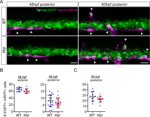
OPCs are specified normally despite reduction in reticulospinal neurons. A, Posterior region of 48-60hpf Tg(olig2:EGFP; sox10:mRFP). EGFP+ mRFP+ cells (OPCs) indicated by arrowheads. At 60hpf some OPCs have migrated dorsally. Scale bar: 20 μm. B-C, Similar number of OPCs in 448μm-long regions of the spinal cord between WT and mutants at 48hpf (B) and 60hpf (C). Data from N = 10 WT and N = 9 mutants (48hpf anterior and 60hpf posterior) and N = 18 WT and N = 20 mutants. Error bars indicate ± SD. |
Expression Data
| Genes: | |
|---|---|
| Fish: | |
| Anatomical Terms: | |
| Stage Range: | Long-pec to Pec-fin |
Expression Detail
Antibody Labeling
Phenotype Data
Phenotype Detail
Acknowledgments
This image is the copyrighted work of the attributed author or publisher, and
ZFIN has permission only to display this image to its users.
Additional permissions should be obtained from the applicable author or publisher of the image.
Full text @ PLoS One