
Fig. 2
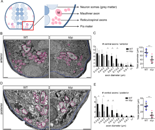
Fewer large-calibre axons in the ventral spinal cord of kbp mutants. A, Diagram of transversal section of spinal cord. Grey matter is in the centre (blue) and white matter or axonal cross-sectional profiles around it (pink). Red box indicate approximate areas shown in B and D. B, D, Transmission electron micrographs of ventral regions of the anterior (B) and posterior (D) spinal cord at 72hpf. Large axons are traced in pink, pia mater in blue. M = Mauthner axon. Scale bar: 1μm. C, E, Distribution of large ventral axons in WT and kbp mutant shows significant reductions in the number of large (reticulospinal) axons in mutants in both the anterior (C) and posterior (E) spinal cord. Graphs on right show that the total perimeter belonging to large axons is significantly reduced in mutants in both regions. Data from N = 5 WT and N = 5 mutant larvae. * indicates P<0.05; ** indicates P<0.01; see text for details. Error bars indicate ± SD. |
| Fish: | |
|---|---|
| Observed In: | |
| Stage: | Protruding-mouth |