
Fig. 3
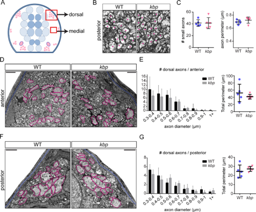
Dorsal and medial axons are not affected in kbp mutants. A, Diagram of hemi-spinal cord transversal section with red boxes indicating dorsal and medial regions shown in B, D and F. B, Transmission electron micrographs of medial region of the posterior spinal cord, with dots indicating small-diameter axons. Scale bar: 0.2μm. C, number and average perimeter of medial axons. D, F, Electron micrographs of dorsal region of the anterior (D) and posterior (F) spinal cord. Large axons are traced in pink, pia mater in blue. Scale bar: 1μm. E, G, Similar distributions of large dorsal axons in WT and kbp mutant anterior (E) and posterior (G) spinal cord. The total perimeter belonging to large axons is similar in WT and mutants. Data from N = 5 WT and N = 5 mutant larvae. Error bars indicate ± SD. |