
Fig. 4
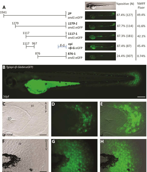
Epi fragment contains an ectodermal enhancer sufficient to recapitulate and1 expression in epithelium of embryonic fin folds. (A) Analysis of truncated 2Pand1 reporter constructs. Constructs were tested for ectodermal enhancer activity. Percentage of eGFP positive embryos during injections, and an average % MFF transient fluorescence coverage are presented on the right (N=Number of total viable injected embryos). Note that the 150 b.p. region 1117-967and1 mimics transient ectodermal enhancer activity of full 2Pand1 element, when tested with the β-globin minimal promoter. Note the reduced percentages for Tg(876and1:eGFP) suggesting the absence of the ectodermal enhancer. (B-H) Tg(epi+β-globin:eGFP) transgenic reporter line at 3 d.p.f. Median (F-H) and pectoral fin fold (C-E) display reporter expression in the ectodermal cells only. Absence of mesenchymal expression is best observed using confocal microscopy. Brightfield (C, F), fluorescence (B, D, G), and brightfield/fluorescence merged images (E, H). ED, Endoskeletal Disc; FF, Fin Fold; A, Anterior; P, Posterior; D, Dorsal; V, Ventral. Scale bars: 200 µm in B; 50 µm in C-H. |
Reprinted from Developmental Biology, 417(1), Lalonde, R.L., Moses, D., Zhang, J., Cornell, N., Ekker, M., Akimenko, M.A., Differential actinodin1 regulation in zebrafish and mouse appendages, 91-103, Copyright (2016) with permission from Elsevier. Full text @ Dev. Biol.