
FIGURE
Fig. S6
Fig. S6
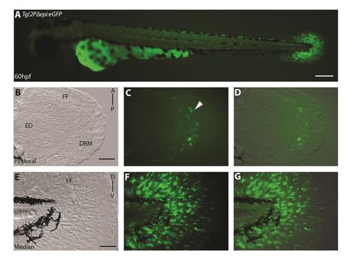
Symmetrical GFP-expressing cells in distal rim mesenchyme at 60 h.p.f. (A-G) 60 h.p.f. in Tg(2PΔepi:eGFP) transgenic reporter line. Mesenchymal expression is present in both the median fin fold (E-G) and the pectoral fin fold (B-D) at 60 h.p.f.. The distal rim mesenchyme of the pectoral fin bud symmetrical reporter expression (C-D). Brightfield (B, E), fluorescence (C-F), and brightfield/fluorescence (D-G) merged images. FF, Fin Fold; ED, Endoskeletal Disc, A, Anterior; P, Posterior; D, Dorsal; V, Ventral. Scale bars: 200µm in A, 50µm in B-G. |
Expression Data
Expression Detail
Antibody Labeling
Phenotype Data
Phenotype Detail
Acknowledgments
This image is the copyrighted work of the attributed author or publisher, and
ZFIN has permission only to display this image to its users.
Additional permissions should be obtained from the applicable author or publisher of the image.
Reprinted from Developmental Biology, 417(1), Lalonde, R.L., Moses, D., Zhang, J., Cornell, N., Ekker, M., Akimenko, M.A., Differential actinodin1 regulation in zebrafish and mouse appendages, 91-103, Copyright (2016) with permission from Elsevier. Full text @ Dev. Biol.