
Fig. S4
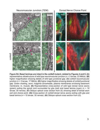
Basal laminae are intact in the col4a5 mutant, related to Figures 4 and 5. (A) representative ultrastructure of wild type neuromuscular junction (n = 3 larvae, 23 NMJs). (B) higher magnification showing details of wild type junctional gap. (C) col4a5 neuromuscular junction (n = 3 larvae, 17 NMJs). (D) higher magnification showing details of col4a5 junctional gap. Green shading, axon; magenta arrowheads, plasma membranes enclosing basement membrane; m, muscle. (E) Representative cross-section of wild type dorsal nerve axons (green) exiting the spinal cord surrounded by glia (red) and basal lamina (cyan) (n = 10 larvae, 30 nerves). (F) Oblique optical cross section from (E) showing detail of dorsal axon exit and choice point. (G) Cross-section of col4a5 dorsal nerve axons exiting with glia and basal lamina (n = 10 larvae, 30 nerves). (H) Oblique optical cross section from (G). |