
Fig. 4
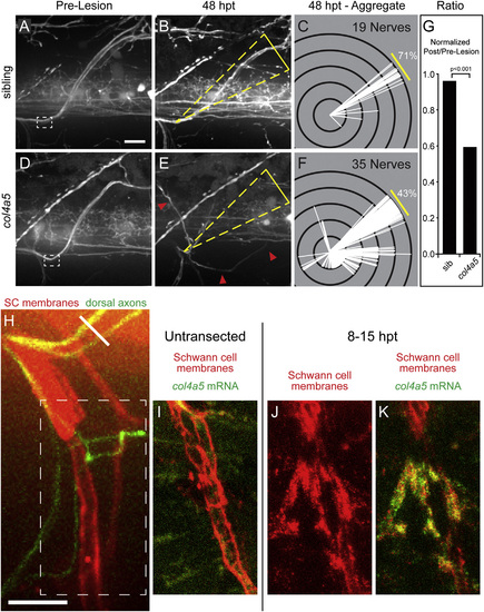
The lh3 Substrate collagen4a5 Is Upregulated after Nerve Transection and Directs Regenerating Dorsal Nerve Axons (A–F) Compared to pre-lesion (A), 48 hpt sibling nerves (B) regenerate to the original outgrowth pathway. Modified Sholl analysis (C; n = 7 larvae, 19 nerves) reveals that the majority of sibling nerves regenerate on the correct path. In contrast, pre-lesion (D) and 48 hpt (E) col4a5 nerve examples and Sholl analysis (F; n = 12 larvae, 35 nerves) show that mutant nerves frequently regrow into aberrant regions of the myotome (yellow triangles, dorsal ROI; red arrowheads, misguided fascicle). (G) These differences were statistically significant after adjusting for developmental dorsal axon patterning in the directionality ratio. (H) Region of transected nerves showing col4a5 mRNA signal in (J) and (K) (oblique white line, transection site; white dashed box, region of nerve shown in I–K). (I) col4a5 in situ hybridization in untransected hemisegments revealed sparse signal (n = 12 larvae, 36/54 nerves). (J and K) 8–15 hr post-transection, col4a5 mRNA (J) was upregulated in Schwann cells (K) ventral and ventrolateral to the transection site (n = same 12 larvae, 52/60 nerves; p < 0.001). All scale bars, 10 µm. |