
FIGURE
Fig. S2
- ID
- ZDB-FIG-151013-15
- Publication
- Ernest et al., 2015 - A genomic region encompassing a newly identified exon provides enhancing activity sufficient for normal myo7aa expression in zebrafish sensory hair cells
- Other Figures
- All Figure Page
- Back to All Figure Page
Fig. S2
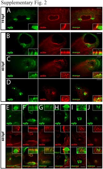
Transgene expression is localized to sensory hair cells of the inner ear and lateral line - Morphological assessment. In order to determine the identity of the cells expressing EGFP reporter gene in the inner ear and lateral line, co-labeling of EGFP (immunohistochemistry) and actin (TRITC-phalloidin) was performed on fixed embryos. Embryos from Tg(B1B2-761bp:EGFP) transgenic line were analyzed at 19hpf (A), 40hpf (B-D) and 60 hpf (E-J). For each developmental stage, EGFP (green) and actin (red) were visualized on an upright spinning disk microscope ; a merge of the two signals is also represented. Inserts show higher magnification of hair cells from the observed sensory patch: tether cells (devoid of hair bundle at 19 hpf) (A), and differentiated hair cells from the anterior maculae (B,F), the posterior maculae (C,G), anterior cristae (E), posterior cristae (H), and neuromasts (D,I,J). Images are z-projections of stacks allowing to visualize together hair cell body and bundle within one sensory patch. Neuromasts are shown entirely (D,I) or only in their most apical part (J). Scale bar = 25 µm. |
Expression Data
Expression Detail
Antibody Labeling
Phenotype Data
Phenotype Detail
Acknowledgments
This image is the copyrighted work of the attributed author or publisher, and
ZFIN has permission only to display this image to its users.
Additional permissions should be obtained from the applicable author or publisher of the image.
Full text @ Dev. Neurobiol.