
Fig. 2
- ID
- ZDB-FIG-151013-8
- Publication
- Ernest et al., 2015 - A genomic region encompassing a newly identified exon provides enhancing activity sufficient for normal myo7aa expression in zebrafish sensory hair cells
- Other Figures
- All Figure Page
- Back to All Figure Page
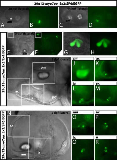
DNA sequences driving expression in hair cells are located within Intron 2–3. Transient expression of EGFP placed under the control of a BAC containing various portions of myo7aa gene. Upper panel shows the lateral view of the otic vesicle of a 28 hpf (A,B) and a 44 hpf (C,D) embryo injected with recombinant BAC 29o13-myo7aa_Ex2/SP6:EGFP. A and C are bright field images of the region observed by epifluorescence microscopy in B and D. EGFP is observed in sensory hair cells of the anterior maculae (B,D). Central panel shows the lateral view (E–H) of a 3 dpf embryo and the dorsal view (J–M) of a 5 dpf embryo injected with recombinant BAC 29o13-myo7aa_Ex3/Ex4:EGFP. Transient expression of EGFP was monitored using a Zeiss epifluorescence microscope. E shows the bright field image of the region located directly posterior to the eye; F shows the fluorescent signal of the exact same field observed in panel E. Panel G (superimposition of bright field and fluorescent images) and H (fluorescence only) are high magnification views of the neuromast boxed in panel E and F. (I) model dorsal view of the left ear of a 5 dpf zebrafish on which the different sensory patches are boxed to facilitate identification and recognition of the sites where fluorescent signal is observed in panels J–M. EGFP is observed in sensory hair cells of the posterior maculae (J) and the three cristae (K–M). Lower panel shows the lateral view of a 5 dpf embryo injected with recombinant BAC 29o13-myo7aa_Ex3/SP6:EGFP. (N) model lateral view of the left ear of a 5 dpf zebrafish on which the different sensory patches are boxed to facilitate identification and recognition of the sites where fluorescent signal is observed in panels O–R. EGFP fluorescence is observed in sensory hair cells of the anterior maculae (O), of the anterior and lateral cristae (P,Q) and in a head neuromast (R). Live embryos were anesthetized with tricaine and mounted in agarose before imaging on a Zeiss epifluorescence microscope. All images are single z-axis sections. am: anterior maculae; pm: posterior maculae; ac: anterior cristae; lc: lateral cristae; pc: posterior cristae; n : neuromast. |