
Fig. S3
- ID
- ZDB-FIG-151013-16
- Publication
- Ernest et al., 2015 - A genomic region encompassing a newly identified exon provides enhancing activity sufficient for normal myo7aa expression in zebrafish sensory hair cells
- Other Figures
- All Figure Page
- Back to All Figure Page
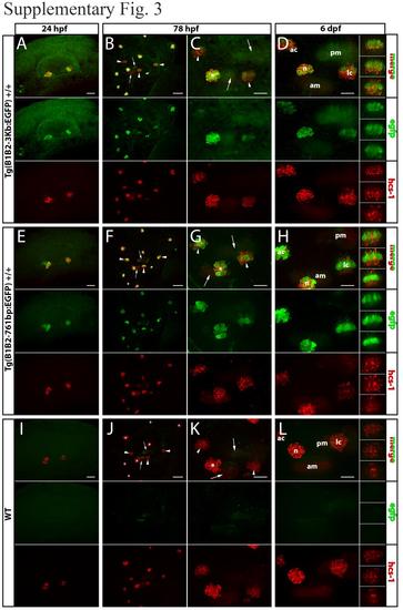
Transgene expression is localized to sensory hair cells of the inner ear and lateral line - Molecular assessment. The identity of the cells expressing EGFP reporter gene within in the inner ear and lateral line was also investigated by double immunofluorescence staining of EGFP and HCS-1, a hair cell-specific antigen, performed on fixed embryos. Embryos from Tg(B1B2-3Kb:EGFP) (A-D) and Tg(B1B2-761bp:EGFP) (E-H) transgenic lines as well as wild-type embryos (I-L) were analyzed at 24 hpf (A,E,I), 78 hpf (B-C,FG, J-K) and 6 dpf (D,H,L). For each developmental stage, EGFP (green) and HCS-1 (red) were visualized on an upright spinning disk microscope. Within each panel a merge of the two signals is represented on top, EGFP labeling in the central portion and HCS-1 labeling at the bottom. EGFP and HCS-1 co-localize in differentiated hair cells of the different sensory patches of the inner ear and of neuromasts. (B,C,F,G,J,K) arrow = anterior or posterior maculae ; arrowhead = anterior, lateral or posterior cristae ; * = neuromast. (D,H,L) am = anterior maculae ; pm = posterior maculae ; ac = anterior cristae ; lc = lateral cristae ; n = neuromast. Scale bar: 20 µm in all panels except for panels B,F,J (50 µm). Images are z-axis projections of z-stacks except for insets on the right hand side of panels D,H,L which are single sections of the lateral cristae. Within each panel, these 3 single sections of the lateral cristae imaged from lateral (upper section) to medial (lower section) are separated 7 µm away from each other. |