
Fig. 7
- ID
- ZDB-FIG-151013-13
- Publication
- Ernest et al., 2015 - A genomic region encompassing a newly identified exon provides enhancing activity sufficient for normal myo7aa expression in zebrafish sensory hair cells
- Other Figures
- All Figure Page
- Back to All Figure Page
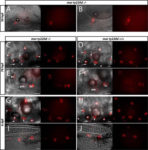
Compensation of mariner ty220d -/- mutation by expression of mCherry-Myo7aa fusion protein under the control of B1B2mB-761bp DNA element. Lateral views of homozygous transgenic Tg(B1B2mB-761bp:mCherry-Myo7aa) zebrafish embryos. (A,C,E,G,I) homozygous mar ty220d/ larvae; (B,D,F,H,J) heterozygous mar ty220d+/- larvae. Live embryos were anesthetized with tricaine and mounted in agarose before imaging on a Leica spinning disk microscope. Embryonic stages imaged were: 30 hpf (A,B), 78 hpf (C–F), 6 dpf (G–J). All panels comprise the superimposition of bright field with fluorescence image (left part) and the fluorescence image (right part) of the exact same area of the embryo. (C,D,G,H) arrow = anterior or posterior maculae; arrowhead = anterior, lateral or posterior cristae; * = neuromast. (E,F) am = anterior maculae; ac = anterior cristae; lc = lateral cristae. (A,B) single z-axis section; (C,J) z-axis projection. Scale bar: 20 µm in all panels except for panels B (10 µm). |