
Fig. S7
- ID
- ZDB-FIG-150616-16
- Publication
- Dalgin et al., 2015 - Differential levels of Neurod establish zebrafish endocrine pancreas cell fates
- Other Figures
- All Figure Page
- Back to All Figure Page
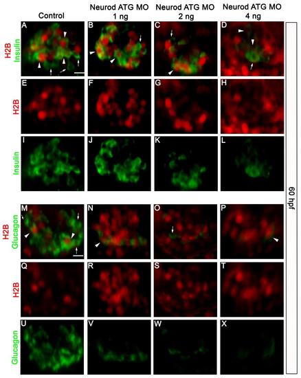
Label retaining cell analysis confirmed that early (dorsal) and late (ventral) born endocrine cells are similarly affected in Neurod morphants. Confocal images (merged z-stacks) of representative 60 hpf Tg(neurod:EGFP) embryos injected with H2B-RFP mRNA (red). GFP channel is not shown and hormone markers are false colored in green for presentation. Whole mount immunolabeling for insulin (green, |
Reprinted from Developmental Biology, 402(1), Dalgin, G., Prince, V.E., Differential levels of Neurod establish zebrafish endocrine pancreas cell fates, 81-97, Copyright (2015) with permission from Elsevier. Full text @ Dev. Biol.