|
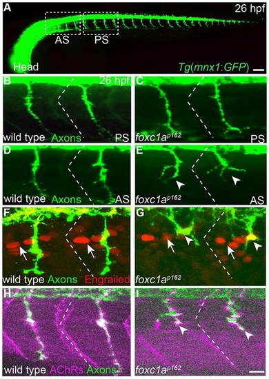
foxc1a guides primary motor axon selectively in anterior somite segments. (A) Lateral (composite) view of a 26-h-old Tg (mnx1:GFP) embryo expressing GFP in all motor neurons. White dashed boxes mark anterior somite segments (AS) and posterior segments (PS) considered in this study. Compared with wild type (B,D), foxc1a mutants (C,E) exhibit motor axon guidance defects selectively in anterior but not posterior somitic segments. (F-I) Somite polarity (F,G), as revealed by the localization of Engrailed-positive nuclei (arrows) towards the anterior somite boundary (dashed lines), and muscle differentiation (H,I), as revealed by the apposition of muscle AChRs with axons to form en passant synapses are unaffected in foxc1a mutants. Arrowheads indicate stalled and branched axons. Scale bars: 50µm in A; 10µm in B-I.
|