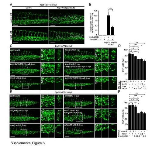
The Bmp-Arhgef9b Signaling Regulates CVP Formation through Fmnl3. Related to Figure 6. (A) Projection view of confocal z-stack images of the caudal regions of 48 hpf Tg(fli1:GFP) embryos injected with 2.5 ng control MO (upper panel) or 2.5 ng fmnl3 MO (lower panel) together without (Control) or with hsp70l:bmp2b-FLAG Tol2 plasmid. (B) The area covered by ectopic venous vessels as observed in A was quantified and expressed as percentages relative to that observed in the embryos injected with both control MO and hsp70l:bmp2b-FLAG plasmid. Data are shown as mean ± s.e.m. (each sample [n=12]). (C) Projection view of confocal z-stack images of the caudal regions of 33 hpf Tg(fli1:GFP) embryos injected with control MO, both bmpr2a and bmp2rb MOs, and fmnl3 MO as indicated in the upper left corner of each image are shown, as in Figure 1G. The amounts of injected MOs are also indicated in parentheses after the name of MO. (D) The width of CVP as observed in C was quantified, as in Figure 1H (n≥5). (E) Projection view of confocal z-stack images of the caudal regions of 33 hpf Tg(fli1:GFP) embryos injected with control MO, arhgef9b MO and fmnl3 MO as indicated in the upper left corner of each image are shown, as in Figure 1G. The amounts of injected MOs (per embryo) are also indicated in parentheses after the name of MO. (F) The width of CVP as observed in E was quantified, as in Figure 1H (n≥5). Scale bars, 100 µm (A, C and E). *p<0.05, **p<0.01, ***p<0.001. n.s., no significance.
|
