|
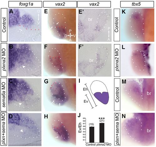
Sema6a/Plxna2 are necessary for proper regionalization of the eye vesicle. Twelve-somite control eyes (A,E,E2,K,M), and plxna2 (B,F,F2,L), sema6a (C,G) and sema6a/plxna2 double (D,H,N) morphant eyes processed with riboprobes for foxg1a (A-D), vax2 (E-H; posterior extent of expression indicated by red dots) and tbx5 (K-N). (A-D) Transverse sections. (E-H,K,L) Lateral views. (M,N) Dorsal views. A/P orientation in E applies. (A-D) foxg1a label is present ventrally in morphants (arrowheads) but not in control (arrow). In E-H, arrows indicate in situ label showing through from the opposite eye vesicle. Mesenchyme (*) is stuck to the eye vesicle in L. (E2,F2) Coronal sections show vax2 mRNA expansion into the posterior eye vesicle in a plxna2 morphant. (I,J) Analysis method (I) and the ratio of vax2 domain height (Ev) normalized to eye vesicle height (Eh) (J). ***P<0.001, unpaired Student′s t-test. Numbers of embryos in brackets (n=2); error bars indicate s.e.m.
|