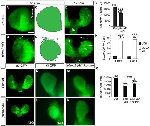
Plxna2 is required for eye tissue cohesion. (A-F) Dorsal confocal images of (A,B) 20- and (E,F) 12-somite rx3:GFP transgenic eye vesicles (e) in control (A,E) and plxna2 morphant (B,F). Scale bar: 50μm in A,B; 100μm in E,F. GFP+ cells are seen ectopic to (arrowheads), emerging from (arrows) and bridging (red arrow, F) the eye vesicles. hy, hypothalamus. Schematics highlight ectopic GFP+ cells in morphants (D), but not in control (C). (G,H) Mean rx3:GFP eye vesicle area (dorsal) (G) and the numbers of ectopic eye cells (H). n=3. Error bars indicate s.e.m. ***P<0.001 unpaired Student′s t-test. (I,J) Dorsal views of 14-somite rx3:GFP control (I) and ATG plxna2 morphant (J). (K-N) Dorsosagittal views of 14-somite rx3:GFP control (K) and e3i3 plxna2 morphant (L), and two embryos co-injected with human PLXNA2 mRNA and e3i3 plxna2 MO (M,N). (O) Quantitation of the dorsosagittal GFP+ eye vesicle area in control, e3i3 plxna2 morphants and e3i3 morphants injected with human PLXNA2 mRNA. The latter have significantly larger eye vesicles than plxna2 morphants, although their eyes are still smaller than control (P<0.001) and some ectopic cells are evident. n=3. Error bars indicate s.e.m., ***P<0.001, one-way ANOVA, Newman-Keuls test.
|
