
Fig. S3
- ID
- ZDB-FIG-140702-52
- Publication
- Ebert et al., 2014 - Sema6a and Plxna2 mediate spatially regulated repulsion within the developing eye to promote eye vesicle cohesion
- Other Figures
- All Figure Page
- Back to All Figure Page
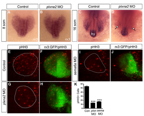
Reduced proliferation in sema6a and plxna2 morphant eyes. (A-D) Dorsal views of wholemount control (A,C) and plxna2 morphants (B,D) processed for rx3 mRNA in situ hybridization at 8 (A,B) and 16 (C,D) somites. rx3 is downregulated by 16 somites in a plxna2 morphant, specifically in the eye (arrows). (E-J) Dorsal views of GFP+ eye vesicles immunostained with anti-pHH3 (red) in control (E,F), plxna2 (G,H), and sema6a (I,J) morphant at 20 somites. (K) Quantitation of the numbers of pHH3+ cells within the eye vesicles of control and morphant embryos. N=2. Error bars represent s.e.m (***, P<0.001; One way ANOVA, Dunnett’s test). |