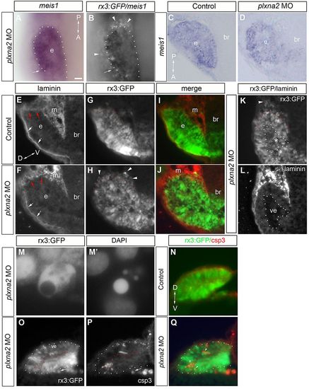
plxna2 morphant cells leave the eye epithelium and undergo apoptosis. (A-D) Lateral whole-mount view (A,B) and coronal sections (C,D) of meis1 label of the eye vesicle (e) of a control (C) and a plxna2 morphant (A,B,D). Ectopic GFP immunoreactive cells (arrowheads) are indicated, as well as an ectopic meis1+ cell (arrow). (E-L) Transverse sections of 12-somite rx3:GFP control (E,G,I) or plxna2 morphant eye vesicles (F,H,J,K,L) immunolabeled with an anti-laminin antibody (E,F,L). The basal lamina (white arrows) is present in both sets of embryos, but is not complete (red arrows). GFP+ cells are found ectopic to the eye vesicle (arrowheads), and within (asterisk) the ventricle (outlined; ve), of the plxna2 morphant (K,L). (M,M2) A GFP+ cell in a plxna2 morphant sits at the vesicle edge (M) with a crenated nucleus (DAPI; M2). (N-Q) Anti-activated caspase 3 immunolabeling (N,P,Q) of transverse sections of control (N) and plxna2 morphant (O-Q; vesicle outlined by dots). Merge with GFP epifluorescence (N,Q). Apoptotic GFP+ cells (arrowheads; Q) are present in the eye vesicle, ventricle and mesenchyme (arrow; P) of the morphant. Scale bar: 20μm for A-L. br, brain; m, mesenchyme.
|
