
Fig. 5
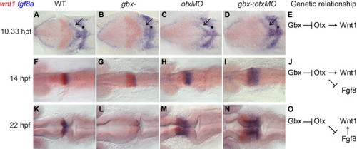
Changing requirements for wnt1 and fgf8 expression leads to rescue of a wnt1–fgf8 boundary in gbx-;otxMO embryos. RNA in situ hybridizations with wnt1 (red) and fgf8a (blue) at the stages shown on the left; genotypes indicated at the top. Dorsal views with anterior to the left. (A–D, F–I, and K–N) At 10.33 hpf (1–3 somites) in WT (A), wnt1 (red) and fgf8a (blue) are expressed in broad domains anterior and posterior to the presumptive MHB respectively (black arrow indicates the fgf8a domain in the anterior hindbrain). fgf8a is also expressed in hindbrain rhombomere 4 (black dot). In gbx-embryos wnt1 is initially expanded (B and G) but subsequently lost (L). Conversely, in otxMO and gbx-;otxMO, wnt1 expression is initially reduced (C and D) but subsequently recovers while the fgf8a domain expands (H, I, M, and N). (E, J, and O) Genetic pathways indicate the decreasing dependence of Wnt1 expression on Otx in the midbrain and its increasing dependence on Fgf8 signaling from r1. |
| Genes: | |
|---|---|
| Fish: | |
| Knockdown Reagents: | |
| Anatomical Term: | |
| Stage Range: | 1-4 somites to 26+ somites |
Reprinted from Developmental Biology, 386(1), Su, C.Y., Kemp, H.A., and Moens, C.B., Cerebellar development in the absence of Gbx function in zebrafish, 181-90, Copyright (2014) with permission from Elsevier. Full text @ Dev. Biol.