FIGURE

Fig. S3

ID
ZDB-FIG-140306-28
Publication
Su et al., 2014 - Cerebellar development in the absence of Gbx function in zebrafish
Other Figures
All Figure Page
Back to All Figure Page
Fig. S3

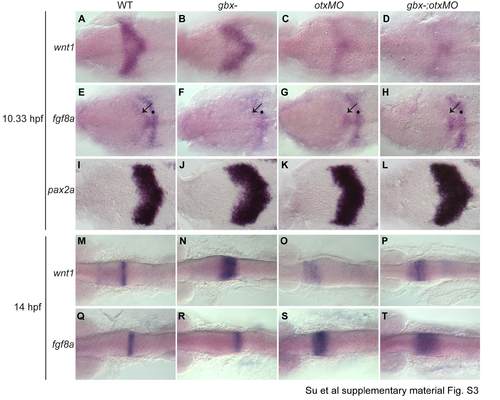
Single color in situs in support of Fig. 5.
RNA in situ hybridizations with wnt1, fgf8a, and pax2a at the stages shown on the left; genotypes indicated at the top. Dorsal views with anterior to the left. (A-L) 10.33 hpf embryos (1-3 somite stage). wnt1 (A-D) is expressed in the presumptive midbrain and is reduced in otxMO and gbx-;otxMO. fgf8a (EH) is expressed in presumptive r1 (black arrows) and r4 (black dots). pax2a (I-L) is expressed in a broad domain encompassing the MHB. The onset of fgf8a and pax2a expression is largely unaffected in mutants compared to WT. (M-T) At 14 hpf, wnt1 expression is still expanded in gbx- and reduced in otxMO and gbx-;otxMO (N-P), while fgf8a expression is progressively expanding in otxMO and gbx-;otxMO (S,T).

Expression Data

Expression Detail
Antibody Labeling
Phenotype Data

Phenotype Detail
Acknowledgments
This image is the copyrighted work of the attributed author or publisher, and ZFIN has permission only to display this image to its users. Additional permissions should be obtained from the applicable author or publisher of the image.

Reprinted from Developmental Biology, 386(1), Su, C.Y., Kemp, H.A., and Moens, C.B., Cerebellar development in the absence of Gbx function in zebrafish, 181-90, Copyright (2014) with permission from Elsevier. Full text @ Dev. Biol.