FIGURE

Fig. S4

ID
ZDB-FIG-140306-29
Publication
Su et al., 2014 - Cerebellar development in the absence of Gbx function in zebrafish
Other Figures
All Figure Page
Back to All Figure Page
Fig. S4

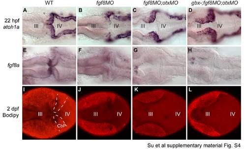
Fgf signaling is required for rescue of the MHB program but not URL specification in gbx- ;otxMO embryos; data in support of Fig. 6.
RNA in situ hybridizations at 22 hpf with the genes indicated on the left (A-H), and Bodipy staining at 2 dpf (I-L). Genotypes are indicated at the top. Embryos are mounted in dorsal view with anterior to the left. (A-D) atoh1a in the URL is absent in fgf8MO (B), but is rescued in fgf8MO;otxMO (C) and even more extensively rescued in gbx-;fgf8MO;otxMO (D). The midbrain (III) and hindbrain (IV) ventricles inflate without forming a sharp isthmic constriction when Fgf8 is knocked down. (E-H) fgf8a expression in anterior r1 is lost in fgf8a morphants (F) and is not rescued by Otx knock-down (G,H). (I-L) 2 dpf embryos stained with CellTraceTM Bodipy, imaged live. Doted lines in WT (I) show the cerebellar primordium (CbP). MHB morphogenesis is disrupted (the III and IV ventricles fuse into a single ventricle) in fgf8MO (J), and is not rescued by knock-down of Otx in otxMO (K) or gbx-;otxMO (L) in spite of prior rescue of cerebellar primordium morphogenesis and URL specification.

Expression Data

Expression Detail
Antibody Labeling
Phenotype Data

Phenotype Detail
Acknowledgments
This image is the copyrighted work of the attributed author or publisher, and ZFIN has permission only to display this image to its users. Additional permissions should be obtained from the applicable author or publisher of the image.

Reprinted from Developmental Biology, 386(1), Su, C.Y., Kemp, H.A., and Moens, C.B., Cerebellar development in the absence of Gbx function in zebrafish, 181-90, Copyright (2014) with permission from Elsevier. Full text @ Dev. Biol.