
Fig. S3
- ID
- ZDB-FIG-130828-30
- Publication
- Gupta et al., 2013 - An Injury-Responsive Gata4 Program Shapes the Zebrafish Cardiac Ventricle
- Other Figures
- All Figure Page
- Back to All Figure Page
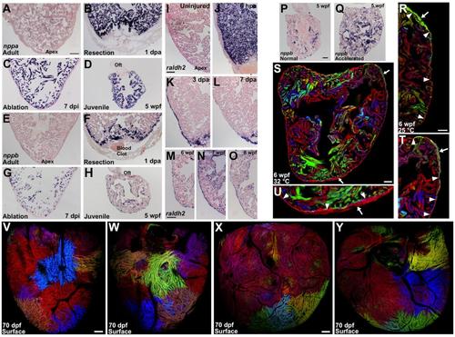
Injury and Stress Markers Are Expressed During Cortical Layer Formation, and Low-Density or High-Temperature Growth Experiments with cmlc2:CreER; priZm Animals (A) Section of an adult ventricle stained by in situ hybridization for nppa, indicating low but detectable expression. (B and C) By 1 day after apical resection (B) (dpa) or 7 days after diffuse genetic muscle ablation (dpi) (C), nppa is strongly induced in cardiomyocytes. (D) nppa expression is detected throughout the 5 weeks post-fertilization (wpf) ventricle (n = 8-14). (E) Section of an adult ventricle stained by in situ hybridization for nppb, indicating no expression. (F and G) By 1 day post amputation (dpa) (F) or 7 days after diffuse genetic muscle ablation (dpi) (G), nppb is induced in cardiomyocytes. (H) nppb expression is detected throughout the 5 weeks post-fertilization (wpf) ventricle (n = 8-14). (I-L) Sections of adult ventricles stained by in situ hybridization for raldh2. There is little or no detectable expression within an uninjured ventricle (I). After partial ventricular resection, raldh2 is induced organ-wide in the endocardium by 6 hours post-amputation (hpa) (J) and organ-wide in the epicardium by 3 dpa (K). Expression in these cell layers localizes to the injury site by 7 dpa (L). (M-O) During juvenile growth, raldh2 is detected in both epicardial and endocardial tissues (n = 8-20). Oft, outflow tract. (P and Q) Sections of 5 wpf ventricles stained by in situ hybridization for nppb, indicating higher expression and suggesting greater biomechanical stress in animals raised under low-density conditions that accelerate growth (n = 8-10). (R) Juvenile animals grown at 25°C have an average of 1.1 ± 0.4 clones per heart at 6 wpf (n = 8). Here, a green cortical clone (arrow) can be seen at the base of heart external to the primordial muscle (arrowheads). (S) Juvenile animals grown at 32°C have an average of 4.0 ± 0.6 clones per heart at 6 wpf (n = 8). Here, two cortical clones (arrows) can be seen at the base (gray) and the apex (red). (T and U) Higher magnification of the two cortical clones from (S). The primordial layer is indicated by arrowheads. (V and W) Animals were labeled at 2 dpf and analyzed at 70 dpf. Ventral (V) and dorsal (W) views of same ventricle in which many small cortical clones are visible. (X and Y) A different ventricle. Many small cortical clones are visible, especially at the base of the ventricle. Scale bars = 100 μm (A-O and V-Y); 50 μm (P-U) |
| Genes: | |
|---|---|
| Fish: | |
| Condition: | |
| Anatomical Term: | |
| Stage Range: | Days 30-44 to Adult |
| Fish: | |
|---|---|
| Condition: | |
| Observed In: | |
| Stage: | Adult |