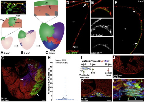
gata4 Marks and Traces Emerging Juvenile Cortical Cardiomyocytes and Regenerating Cardiomyocytes (A) The 30 dpf ventricle comprises an outer primordial layer of single cardiomyocyte thickness. By <6 weeks postfertilization (wpf), inner trabecular cardiomyocytes (Tr) (green) breach the primordial layer (Pr) to establish a cortical cardiomyocyte clone (Cor) at the base of the ventricle. Top: histology section from multicolor clonal analysis, indicating breaching (arrows) of the primordial layer (arrowheads) by a trabecular myofiber (green). (B) During maturation, the initial basal clone (green) expands while other clones emerge on the ventricular surface. (C) Bottom: cortical layer development is completed by adulthood, with a small number of clones contributing to the entire cortical layer. Top: the adult ventricle retains an architecture with three muscle types. (D–F) Tissue sections of 6 (D and E) and 8 (F) weeks postfertilization (wpf) ventricular portions, assessed for tcf21:DsRed2+ epicardial cells (red) and gata4:EGFP+ cardiomyocytes (green). At 6 wpf, a cluster of gata4:EGFP+ surface myocytes is visible at the base of the heart in an area of dense epicardial tissue (arrow in D). By 8 wpf, gata4:EGFP+ cortical myocytes are detected at the ventricular midpoint, with cortical gata4:EGFP expression strongest in cardiomyocytes closer to the apex (arrow) and weaker in basal myocytes (arrowhead) (n = 8–12). Tr, trabeculae; Oft, Outflow tract. (G) gata4:ERCreER; priZm2 animals were pulsed with 4-HT at 6 wpf, and ventricles were analyzed at 90 dpf. Image shows surface muscle from a ventricle in these experiments with arrows indicating large clones. (H) Percentage surface area (SA) occupied by each clone from experiments in (G) (96 total counted clones from nine ventricular halves). (I) Cartoon of lineage-tracing experiments to determine clonal contributions in regenerating gata4+ cardiomyocytes. (J) Section through a ventricle from an uninjured animal 10 days after 4-HT treatment, indicating no recombination in the absence of injury. (K) Section through a ventricular apex at 30 dpa, visualizing multiple colored cardiomyocyte clones in regenerated muscle. Over 15 colors can be distinguished in this 10 µm section of the regenerate. A maximum projection image from confocal slices is shown (n = 6). (L) Higher-magnification view of regenerate shown in (K). Arrows indicate three differently colored multicellular clones. Scale bars represent 100 μm (D–G) and 50 μm (J–L). See also Figure S1 and Movie S1.
|
