
Fig. S1
- ID
- ZDB-FIG-130828-28
- Publication
- Gupta et al., 2013 - An Injury-Responsive Gata4 Program Shapes the Zebrafish Cardiac Ventricle
- Other Figures
- All Figure Page
- Back to All Figure Page
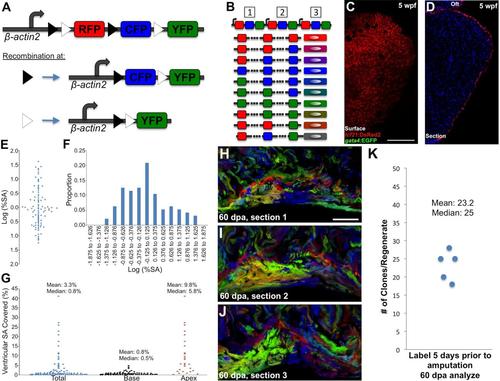
Brainbow Genetics, Early gata4 Expression, Statistical Analysis of gata4:ERCreER; priZm2 Clones, and Clonal Analysis of Regenerating Cardiomyocytes (A) A cartoon of the priZm construct that drives expression of the Brainbow 1.0L cassette [1] with zebrafish β-actin2 regulatory sequences. RFP is the initial reporter that is expressed in the absence of recombination. Recombination at the paired lox2272 (black triangles) or loxP (white triangles) sites results in expression of CFP or YFP, respectively. (B) Tandem insertions of a transgene at a single genetic locus are a common outcome of transgenesis. In the case of 3 transgenes, limited Cre-mediated stochastic recombination upon each Brainbow cassette will result in one of 10 possible outcomes that gives rise to 10 different colors. As the number of Brainbow cassettes increases, so do the possible number of unique outcomes and colors. (C and D) Whole-mount (C) and section images (D) of a 5 wpf ventricle assessed for tcf21:DsRed2+ epicardial cells (red) and gata4:EGFP+ cardiomyocytes (green). gata4+ cardiomyocytes are not detectable. (E) A plot of the log-transformed % surface areas values from Figure 1G. The values have an average of -0.018 ± 0.665 (mean ± standard deviation). A Shapiro-Wilk test found that they differed significantly from a normal distribution, p < 0.03. (F) Distribution of the log-normalized values from (E), indicating an upper tail that appears to represent a separate population of large clones. (G) Percentage surface area (SA) occupied by cardiomyocyte clones generated after 6 wpf labeling in gata4:ERCreER; priZm2 animals. All clone sizes were plotted (total), and then sizes were categorized regionally by location at the base or those that reached past the chamber midpoint (apex) (n = 69 basal and 27 apical clones from 9 ventricular halves). (H-J) Adult cmlc2:CreER; priZm animals were labeled with 4-HT 5 days prior to amputation, and ventricular apices were resected 5 days later. Three serial 30-micron sections through a 60 dpa regenerate (H-J), in which multiple interwoven small clones can be visualized within the regenerate. Clonal representation changes markedly between sections. Dashed lines indicate the approximate plane of resection. (K) Number of clones counted from 3 serial sections of the regenerates in 5 different cmlc2:CreER; priZm ventricles. The number of colors counted in these regenerates likely reaches the limit for this transgenic line. Scale bar = 100 μm (C and D); 50 μm (H-J) |