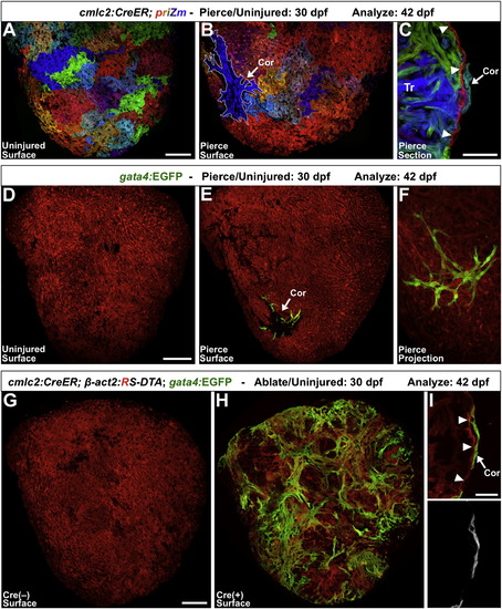
Experimental Injuries Stimulate Ectopic Cortical Muscle Formation (A) cmlc2:CreER; priZm zebrafish were labeled at 2 dpf, assessed at 42 dpf, and indicate no signs of cortical cardiomyocytes on the ventral ventricular surface. (B) When this region of the ventricle was pierced with a fine needle at 30 dpf, a small cortical clone (outlined in white; Cor) was detected at 42 dpf at the injury site in nearly half of the animals. (C) Section indicating a cortical clone (arrow) at 42 dpf, in a ventricle that had been pierced at 30 dpf. Arrowheads indicate the single-cardiomyocyte-thick primordial layer. (D and E) 30 dpf priZm; gata4:EGFP ventricles were pierced as in (A)–(C), with β-actin2-expressing cells indicated in red. Uninjured animals had no gata4:EGFP+ cardiomyocytes on the ventral surface of the ventricle at 42 dpf (D). By contrast, approximately half of the animals pierced at 30 dpf displayed a focus of gata4:EGFP+ cortical muscle at the site of trauma (E, arrow). (F) Higher-magnification confocal projection of injury site in (E). (G and H) Partial genetic ablation of cardiac muscle was initiated by brief 4-HT treatment at 30 dpf and analyzed 12 days later. 4-HT-treated Cre() ventricles contained no gata4:EGFP+ surface cardiomyocytes (G). By contrast, the ventricular surfaces of 4-HT-treated Cre(+) animals contained large amounts of gata4:EGFP+ cortical muscle (H). (I) Section indicating that gata4:EGFP+ cortical cardiomyocytes (arrow) are located on the outside of the primordial layer (arrowheads). Scale bars represent 100 μm (A, B, and D–H), 50 μm (C), and 25 μm (I). See also Figure S2.
|
