
FIGURE
Fig. S2
- ID
- ZDB-FIG-130828-29
- Publication
- Gupta et al., 2013 - An Injury-Responsive Gata4 Program Shapes the Zebrafish Cardiac Ventricle
- Other Figures
- All Figure Page
- Back to All Figure Page
Fig. S2
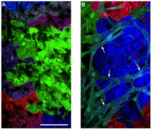
Morphological Differences between Cortical and Primordial Cardiomyocytes (A) An image of a 6 wpf cmlc2:CreER; priZm ventricular surface, from an animal that had been labeled with 4-HT at 2 dpf. This imaged area is not covered with cortical muscle. The cardiomyocytes have limited sarcomeric structure. (B) An image of a 10 wpf cmlc2:CreER; priZm ventricular surface, in which cyan colored cortical myocytes (arrows) are positioned on top of the primordial layer. Cortical myocytes are rod-shaped with clearer sarcomeric organization. Scale bars = 100 μm. |
Expression Data
Expression Detail
Antibody Labeling
Phenotype Data
Phenotype Detail
Acknowledgments
This image is the copyrighted work of the attributed author or publisher, and
ZFIN has permission only to display this image to its users.
Additional permissions should be obtained from the applicable author or publisher of the image.
Full text @ Curr. Biol.