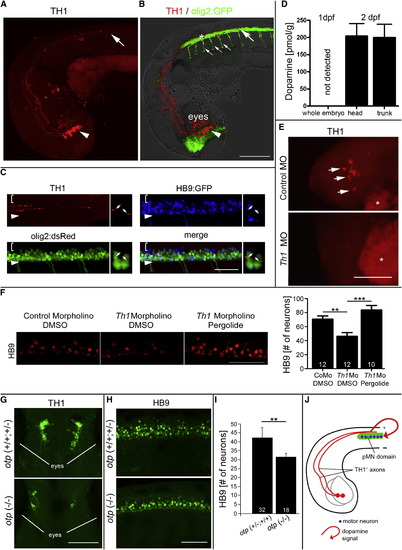
Dopamine from the Brain Promotes Motor Neuron Development (A and B) In a lateral view of (A) TH1 immunohistochemistry only and (B) combined with olig2:GFP fluorescence and light microscopy, diencephalic TH1+ neurons (arrowheads; 33 hpf) and axonal growth cones of their spinal projections (large arrows) are labeled as they grow in close proximity to olig2:GFP+ cells in the ventral spinal cord. Small arrows in (B) indicate motor axons. (C) TH1 labeling reveals close contact of axons (small arrows in coronal views) with pMN progenitors (olig2:dsRed+/HB9:GFP-). Brackets indicate dorsal spinal cord; arrowheads indicate ventral border of spinal cord. (D) Consistent with TH1+ axon growth, HPLC detects dopamine in 48 hpf embryos but not in 24 hpf embryos. (E and F) Injection of a morpholino to th1 leads to (E) loss of TH1 immunoreactivity (indicated by arrows and lateral views of heads; asterisks indicate autofluorescence of the yolk sac) and (F) a significant reduction in the number of HB9+ motor neurons (lateral spinal cord views; 33 hpf), which is fully rescued by pergolide application (drug exposure 24–33 hpf; one-way ANOVA, p = 0.0001, with Bonferroni’s multiple comparison, p < 0.001, p < 0.0001). (G–I) In the otp mutant, fewer dopaminergic neurons are present in the brain, as indicated (G) in dorsal views of heads by TH1 immunohistochemistry, and motor neuron numbers in the spinal cord are reduced, as indicated in (H) lateral views of HB9 immunohistochemistry and (I) quantification thereof. p < 0.01. (J) Model of the spatial relationship of descending dopaminergic axons with the spinal pMN domain. Error bars represent SEM. Scale bars, 200 μm (A and B), 50 μm (C, F, and H), and 100 μm (E and G). See also Figure S1.
|
