
Fig. S5
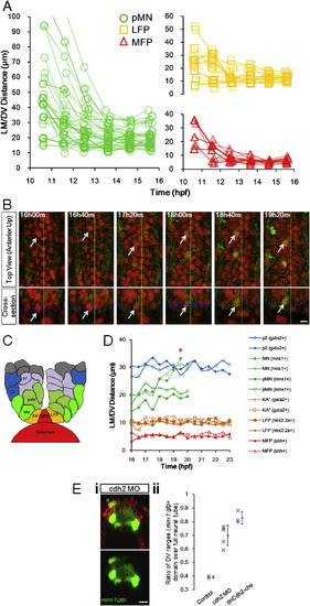
Domain Boundaries Remain Stable after 16 hpf, Cell-type Verifications by Additional Markers, and Additional cdh2 Perturbation Data, Related to Figure 5 (A) Plots by cell type from Figure 5B.(B) LFP cell type confirmation by later stage time-lapse of tg(gata2:gfp) embryos. KA” neurons (gata2:gfp+) are born from the LFP domain among LFP progenitors (nkx2.2a:mgfp+, Figures S1A and S3C; Huang et al., 2012). Note that the tracked LFP cell stays in stable location over time and becomes gata2:gfp+ by 18h40m. All scale bars: 10 μm.(C) Schematic illustration of cell positions in the final pattern. Enlarged and annotated from Figure 1Di.(D) Later stage tracking with various markers. Asterisk: Maturing MNs leave pMN domain and migrate laterally, effectively increasing the LM/DV distance. Neuron movements happen after progenitor domain formation and are distinct from progenitor movements. Cell positions are otherwise stable in this time window indicating stability of established pattern. See also Movie S6.(E) (i).Cdh2 perturbed embryo as in Figure 5F. Example of ectopically localized pMNs that are cherry+ (arrow). Both injected and uninjected (see also Figure 5F) pMNs might become misplaced, suggesting the pattern disruption caused by Cdh2 MO is cell nonautonomous. (ii). Cdh2 perturbed embryos have wider distribution of pMNs. The ratio between the maximum DV range of the mnx1:gfp+ cell cross-sectional distribution over the full length of the neural tube at 24hpf was measured. dnCdh2-che: dominant negative Cdh2-cherry fusion. Blue marks: measurement on one embryo. Red marks: average ratios (±s.d.). |
Reprinted from Cell, 153(3), Xiong, F., Tentner, A.R., Huang, P., Gelas, A., Mosaliganti, K.R., Souhait, L., Rannou, N., Swinburne, I.A., Obholzer, N.D., Cowgill, P.D., Schier, A.F., and Megason, S.G., Specified neural progenitors sort to form sharp domains after noisy shh signaling, 550-561, Copyright (2013) with permission from Elsevier. Full text @ Cell