
Fig. S3
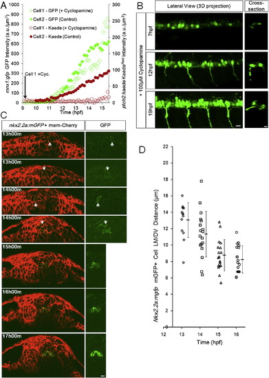
Timing of Specification and Domain Formation of pMNs and LFPs, Related to Figure 3 (A) Specified cells increase marker expression independently from Shh response. 2 representative tracks from a Cyclopamine treated embryo (Cell 1, Cyclopamine added at the start of imaging near 9hpf) and a control embryo (Cell 2, no Cyclopamine). Embryos are double transgenic for ptch2:Kaede and mnx1:gfp. A 405nm laser scan was performed before acquiring each time point to convert Kaede(green) to Kaede(red). 3 additional Cyclopamine tracks and 5 additional control tracks show similar behavior (data not shown).(B) Representative lateral 3D and cross-sectional views of Cyclopamine treated embryos. Early treatment does not fully remove GFP+ (mnx1:gfp) cells, a possible consequence of drug efficiency and/or early pMNs that can be specified in the absence of extracellular Shh (Lewis and Eisen, 2001; Chen et al., 2001). Cross-sections indicate that the specified cells still localize to correct positions. All scale bars: 10 μm. (C) Cross-sectional view of nkx2.2a:mgfp expression at different times. Before 13hpf potential mGFP+ cells could not be uncovered by imaging this transgenic line. At 13 and 14hpf GFP+ cells are found in variable locations and the left-right symmetric distribution of GFP+ cells in the LFP domain is not established. After 15hpf most GFP+ cells line up into the LFP domain on both sides of the triangular MFP cell, this position is indicative of LFP fate after 16hpf.(D) Nkx2.2a:mgfp+ cell distribution over time. Each mark represents the location of one mGFP+ cell at the corresponding time point. Note the narrowing of distribution from 14 to 16hpf and the population average (±s.d.) moving closer to the MFP/notochord. |
Reprinted from Cell, 153(3), Xiong, F., Tentner, A.R., Huang, P., Gelas, A., Mosaliganti, K.R., Souhait, L., Rannou, N., Swinburne, I.A., Obholzer, N.D., Cowgill, P.D., Schier, A.F., and Megason, S.G., Specified neural progenitors sort to form sharp domains after noisy shh signaling, 550-561, Copyright (2013) with permission from Elsevier. Full text @ Cell