
FIGURE
Fig. 6
Fig. 6
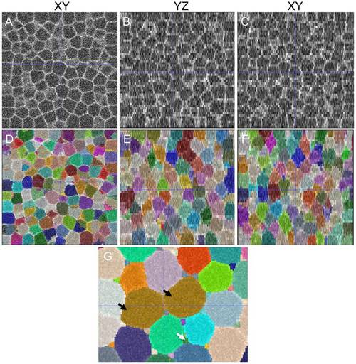
Accurate and highly-sensitive algorithm performance on synthesized 3D membrane images. (A-C) Synthesized cell structures in 3D along XY, YZ and XZ sections with image noise added (Table 2). As in the case of real-world images, the lateral resolution significantly differs from the axial resolution. (D-F) Segmentations overlaid on the raw image with a 50% opacity function. (G) An example of under-segmentation (brown cells, black arrows) and over-segmentation (interstitial fragments, white arrows) in the image. The errors could be filtered out by size criteria. |
Expression Data
Expression Detail
Antibody Labeling
Phenotype Data
Phenotype Detail
Acknowledgments
This image is the copyrighted work of the attributed author or publisher, and
ZFIN has permission only to display this image to its users.
Additional permissions should be obtained from the applicable author or publisher of the image.
Full text @ PLoS Comput. Biol.