
FIGURE
Fig. 5
Fig. 5
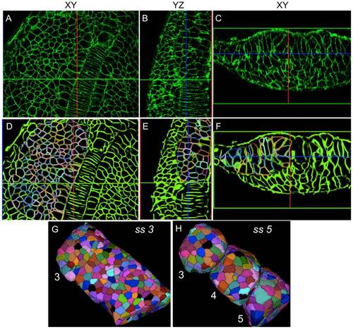
Robust reconstruction and segmentation of cells in the presomitic mesoderm. (A-C) Raw image data showing presomitic mesoderm on 2D image planes (XY,YZ, and XZ) at 3ss. (D-F) Segmentation meshes overlaid on reconstructed membrane images demonstrate excellent localization. Each mesh was randomly colored for visually separating adjacent cells easily. (G,H) 3D rendering of membrane segmentations at 3ss and 5ss. Somites 3, 4 and 5 at 5ss are formed from the presomitic tissue at 3ss by cell sorting and rearrangement. |
Expression Data
Expression Detail
Antibody Labeling
Phenotype Data
Phenotype Detail
Acknowledgments
This image is the copyrighted work of the attributed author or publisher, and
ZFIN has permission only to display this image to its users.
Additional permissions should be obtained from the applicable author or publisher of the image.
Full text @ PLoS Comput. Biol.