
Fig. 2
- ID
- ZDB-FIG-120329-35
- Publication
- Zhu et al., 2012 - Activated ALK Collaborates with MYCN in Neuroblastoma Pathogenesis
- Other Figures
- All Figure Page
- Back to All Figure Page
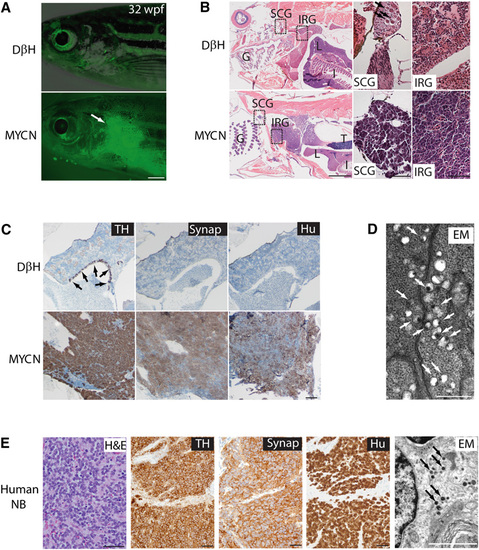
Neuroblastomas Arise in MYCN-Expressing Transgenic Zebrafish (A) Top: DβH fish. Bottom: MYCN fish with EGFP-expressing tumor (arrow) at 32 weeks postfertilization (wpf). Scale bar represents 1 mm. (B) Top: H&E-stained sagittal sections of DβH fish. Boxes indicate the SCG and the IRG, and are magnified in the right panels. Bottom: H&E-stained sagittal sections of MYCN fish with neuroblastic tumors. Boxes indicate the SCG and the IRG and are magnified in the right panels. Arrows indicate SCG neurons. The majority of tumors arise in the IRG of MYCN fish, although as seen in this example, tumor cells in the SCG were occasionally observed in individual fish that also had tumors in the IRG. G, gill; L, liver; I, intestine; IRG, interregnal gland; SCG, superior cervical ganglion; T, testis. Scale bars represent 50 μm. (C) Top: Sagittal sections through the interregnal gland of DβH fish. Chromaffin cells of the interregnal gland express TH (arrows). Bottom: Sagittal sections through the interregnal gland of a MYCN fish with EGFP-expressing tumor. Cells throughout the tumor in the interregnal gland express TH, Synaptophysin (Synap), and Hu. Scale bar represents 100 μm. (D) Electron microscopy (EM) reveals neurosecretory granules in the MYCN-expressing tumors (arrows). Scale bar represents 500 μm. (E) Pathological, immunohistochemical, and ultrastructural analyses of a human neuroblastoma. Arrows point to neurosecretory granules. Scale bars represent 500 μm (left panel), 100 μm (middle panels), and 2 µm (right panel), respectively. See also Figure S2. |
| Gene: | |
|---|---|
| Fish: | |
| Anatomical Term: | |
| Stage: | Adult |
| Fish: | |
|---|---|
| Observed In: | |
| Stage: | Adult |
Reprinted from Cancer Cell, 21(3), Zhu, S., Lee, J.S., Guo, F., Shin, J., Perez-Atayde, A.R., Kutok, J.L., Rodig, S.J., Neuberg, D.S., Helman, D., Feng, H., Stewart, R.A., Wang, W., George, R.E., Kanki, J.P., and Look, A.T., Activated ALK Collaborates with MYCN in Neuroblastoma Pathogenesis, 362-373, Copyright (2012) with permission from Elsevier. Full text @ Cancer Cell