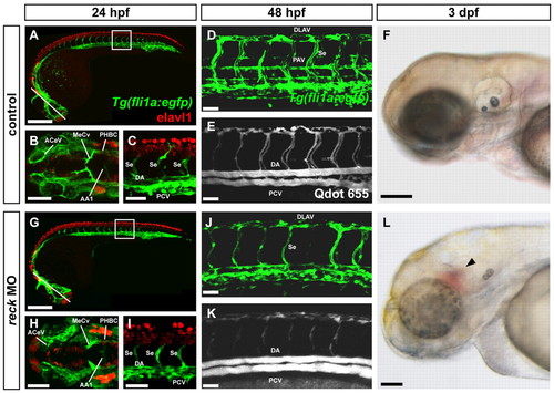
reck-deficient embryos exhibit defects in vascularization. Mock-injected (A-F) and reck MO-injected (G-L) zebrafish embryos. (A,G) Tg(fli1a:egfp) zebrafish embryo (24 hpf) immunostained for Elavl1. Line indicates plane of image in B,H; box indicates area of image in C,I. Scale bars: 250 μm. (B,H) Cranial vasculature. AA1, mandibular arch; ACeV, anterior cerebral vein; MeCV, middle cerebral vein; PHBC, primordial hindbrain channel. Scale bars: 100 μm. (C,I) Trunk vasculature. DA, dorsal aorta; PCV, posterior cardinal vein; Se, intersegmental vessel. Scale bars: 50 μm. All blood vessels form normally in MO-injected animals but are slightly delayed in their extension. (D,J) Tg(fli1a:egfp) embryo (48 hpf). DLAV, dorsal longitudinal anastomotic vessel; PAV, parachordal vessel. The PAV is reduced in size or fails to form entirely in MO-injected embryos. Scale bars: 50 μm. (E,K) Qdot angiogram of the embryo shown in D,J. Although blood flows through the DA and PCV after MO injection, blood flow is somewhat reduced in the DLAV and intersegmental vessels. Scale bars: 50 μm. (F,L) Brightfield image of 3 dpf embryo. Cranial hemorrhage (arrowhead) is visible in a significant fraction of MO-injected embryos. Scale bars: 135 μm.
|
