
Fig. S2
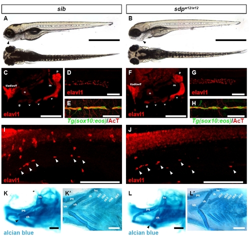
Derivatives of the neural crest other than DRG are largely unperturbed in sdp embryos. (A) Brightfield micrograph of a sibling 4 dpf zebrafish embryo. Scale bar: 500 µm. (B) Brightfield micrograph of a sdpw12/w124 dpf embryo. The sdpw12/w12 jaw is retracted (arrowhead). (C) Cranial ganglia of a sibling 4 dpf embryo as identified by Elavl1 immunostaining. ad, anterodorsal lateral line ganglion; av, anteroventral lateral line ganglion; f, facial ganglion; g, glossopharyngeal ganglion; m, middle lateral line ganglion; o, octaval/statoacoustic ganglion; p, posterior lateral line ganglion; t, trigeminal ganglion; v, vagal ganglia. Scale bar: 100 μm. (F) All cranial ganglia are present in sdpw12/w12 embryos. (D) Enteric nervous system of a sibling 5 dpf embryo as shown by Elavl1 immunostaining. Scale bar: 100 μm. (G) Enteric nervous system of a sdpw12/w125 dpf embryo. The enteric nervous system migrates the full extent of the gut. (E) Lateral line nerve of a sibling 4 dpf embryo stains positive for acetylated tubulin; myelinating glia are marked by the sox10:eos transgene. Scale bar: 50 μm. (H) Lateral line nerve and associated myelinating glia are present in sdpw12/w12 embryos. (I) Sympathetic neurons (arrowheads) in a sibling 5 dpf embryo immunostained for Elavl1. Scale bar: 150 μm. (J) Sympathetic neurons are also present in 5 dpf sdpw12/w12 embryos. Scale bar: 150 μm. (K) Head cartilages of a sibling 4 dpf embryo. Note the position of the ceratohyal cartilage (arrowhead). ch, ceratohyal; hs, hyosymplectic; pq, palatoquadrate. Scale bar: 100 μm. (K2) Branchial arches of a sibling 4 dpf embryo. bh, basihyal; cb, ceratobranchial; ih, interhyal. Scale bar: 100 μm. (L) Head cartilages of a sdpw12/w124 dpf embryo. The ceratohyal cartilage is retracted (arrowhead). Scale bar: 100 μm. (L2) All branchial arches of the sdpw12/w124 dpf embryo are present. Scale bar: 100 μm. |