
Fig. S7
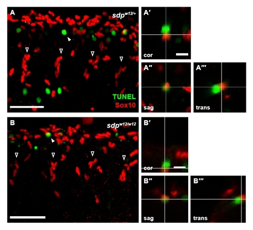
Increased apoptosis is not observed in sdp neural crest. Sdpw12/+ fish were incrossed and the resulting embryos were subjected to Sox10 and TUNEL staining at 30 hpf. Four streams of neural crest cells (open arrowheads) are visible in each image. (A) Embryos heterozygous for sdpw12 exhibit numerous TUNEL+ cells, many of which are in close proximity to Sox10+ cells (filled arrowhead). Scale bar: 50 μm. (A2-A222) Orthogonal projections of confocal stacks were generated to ascertain whether TUNEL and Sox10 staining is colocalized, as evident in the coronal (A2), sagittal (A22) and transverse (A222) projections; although TUNEL+ and Sox10+ cells are adjacent, the markers do not colocalize. Scale bar: 10 μm. (B) Embryos homozygous for sdpw12 exhibit a similar distribution of TUNEL+ and Sox10+ cells. Scale bar: 50 μm. (B2-B222) Again, in coronal (B2), sagittal (B22) and transverse (B222) projections, no colocalization of markers is observed. Scale bar: 10 μm. |