
Fig. 4
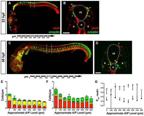
reck expression in neural crest cells. (A) Whole-mount zebrafish embryo (22 hpf). Dashed lines indicate the plane of section shown in B. (B) reck+ cells (red arrowheads) are present in both the developing vasculature and presumptive neural crest. A small number of crestin+ cells are also visible (green arrowhead). Most crestin+ cells are also reck+ (yellow arrowheads). Scale bar: 25 μm. (C) Whole-mount embryo (30 hpf). Dashed lines indicate the plane of the section shown in D. (D) Many reck+ cells (red arrowheads) are present in the developing vasculature. Many crestin+ cells (green arrowheads) are now apparent with few crestin+/reck+ cells (yellow arrowhead), suggesting the two markers have resolved into different populations. Scale bar: 25 μm. (E,F) Cell counts from serial sections of 22 hpf (E) and 30 hpf (F) embryos. Error bars represent s.d. Green, crestin+ cells; yellow, crestin+/reck+ cells; red, reck+ cells. (G) crestin+/reck+ cells as a proportion of all cells expressing crestin for 22 hpf (white circles) and 30 hpf (black circles) embryos. In most segments we observe significant segregation of crestin and reck expression. Error bars represent 95% confidence interval. n, notochord; nt, neural tube; s, somite. |
| Genes: | |
|---|---|
| Fish: | |
| Anatomical Terms: | |
| Stage Range: | 26+ somites to Prim-15 |