
Fig. 3
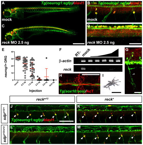
Phenocopy of sdp with reck MO and rescue by expression of wild-type reck. (A-D) Mock injected (A,B) and MO-injected (C,D) 3 dpf Tg(neurog1:egfp) embryos immunostained for Elavl1. Scale bar: 500 μm in A,C; 50 μm in B,D. (E) neurog1+ DRG at 3 dpf in embryos injected with indicated doses of reck MO. Error bars represent s.d. (F) Loss of correctly spliced reck transcript with reck MO. RT–, negative control reaction without reverse transcriptase. (G-I) Tg(sox10:eos) embryo (3 dpf) injected with 1.5 ng reck MO and immunostained for Elavl1. (G) Crest-derived Schwann glia (empty arrowhead) are retained. Scale bar: 25 μm. (H) Crest-derived lateral line glia are retained. Scale bar: 50 μm. (I) Crest-derived pigment cells (melanophore shown) are retained. Scale bar: 25 μm. (J-M) Injection of 200 pg sdpw12 reck mRNA has no effect on sdpw12/+ (J) or sdpw12/w12 (L) fish. Injection of wt reck mRNA has no effect on sdpw12/+ fish (K) but rescues DRG formation in sdpw12/w12 fish (M). Scale bar: 100 μm. Arrowheads indicate DRG neurons. |
| Genes: | |
|---|---|
| Antibody: | |
| Fish: | |
| Knockdown Reagent: | |
| Anatomical Terms: | |
| Stage: | Protruding-mouth |