
Fig. 1
- ID
- ZDB-FIG-120201-39
- Publication
- Tao et al., 2011 - Zebrafish prox1b Mutants Develop a Lymphatic Vasculature, and prox1b Does Not Specifically Mark Lymphatic Endothelial Cells
- Other Figures
- All Figure Page
- Back to All Figure Page
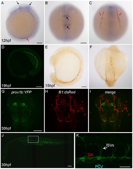
Prox1b is expressed in the endothelial cells and the central nervous system of the head. (A–C) shows prox1b transcript expression by whole mount in situ hybridization in wild-type embryos, at 12 hpf. Black arrows point to prox1b expression in the head; red arrows indicate prox1b expression in lateral plate mesoderm. Confocal image (D) shows YFP expression in a prox1b BAC:YFP embryo at 19 hpf stage. (E) and (F) show YFP expression, enhanced by DAB immunostaining, is detected in prox1b BAC:YFP embryos in migrating angioblasts at 15 hpf. (G–I) shows prox1b:YFP expression in the head region of a prox1b BAC:YFP, fli1:DsRed embryo. Note overlapping (endothelial cells) and non-overlapping expression domains. (J) shows prox1b:YFP expression in the trunk vasculature. (K) shows enlarged view of the boxed area in (J). Scale bars represent 50 μm in (K), and 100 μm in other figures. |
| Genes: | |
|---|---|
| Fish: | |
| Anatomical Terms: | |
| Stage Range: | 5-9 somites to Prim-15 |