
Fig. 4
- ID
- ZDB-FIG-120201-42
- Publication
- Tao et al., 2011 - Zebrafish prox1b Mutants Develop a Lymphatic Vasculature, and prox1b Does Not Specifically Mark Lymphatic Endothelial Cells
- Other Figures
- All Figure Page
- Back to All Figure Page
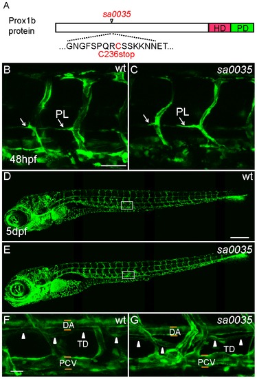
The lymphatic development of homozygous prox1bsa0035 mutants appears normal. (A) Schematic representation of the Prox1b protein, with the position of the prox1bsa0035 allele indicated. The homeodomain region (HD) is shown in red, the Prospero domain (PD) in green. The predicted stop mutation occurs at C236 in prox1bsa0035. (B) and (C) show vascular structures in the trunk region of wild-type (wt, B) and homozygous prox1bsa0035 mutant embryos (C) in fli1:GFP background. The white arrows indicate PLs. (D) and (E) show whole embryo lateral view images of 5-day wt (D) and homozygous prox1bsa0035 mutant embryos (E). (F) and (G) show enlarged views of the boxed areas in (D) and (E). The white arrowheads indicate the presence of TD in both control (F) and homozygous prox1bsa0035 embryos (G). Scale bars represent 50 μm in (B), 250 μm in (D) and 25 μm in (F). |
| Gene: | |
|---|---|
| Fish: | |
| Anatomical Term: | |
| Stage: | Day 5 |
| Fish: | |
|---|---|
| Observed In: | |
| Stage Range: | Long-pec to Day 5 |