
Fig. S1
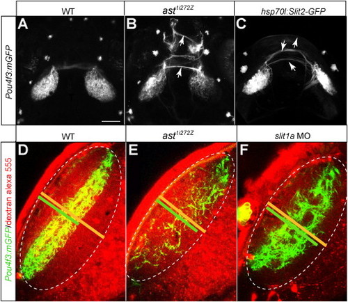
RGC Axon Projections Require Slit/Robo Signaling, Related to Figure 2(A–C) Projections of 6 dpf Pou4f3:mGFP WT (A), astti272z (B), and hsp70l:Slit2-GFP fish (C). Heat shock was applied at 76 hpf.(D–F) Optical sections of tectum of 5 dpf Pou4f3:mGFP fish. Arrows point at RGC axons aberrantly crossing midline. Pou4f3:mGFP-labeled RGC axons occupy about 60% of the depth of the tectal neuropil (D). In both astti272z mutant (E) and slit1a MO (F), Pou4f3:mGFP-labeled RGC axons are more dispersed in the tectal neuropil. Yellow lines in D to F indicate the depth of the tectal neuropil. Green lines in D to F indicate the depth of Pou4f3:mGFP expressing RGC axons. |
Reprinted from Cell, 146(1), Xiao, T., Staub, W., Robles, E., Gosse, N.J., Cole, G.J., and Baier, H., Assembly of Lamina-Specific Neuronal Connections by Slit Bound to Type IV Collagen, 164-176, Copyright (2011) with permission from Elsevier. Full text @ Cell