
Fig. S3
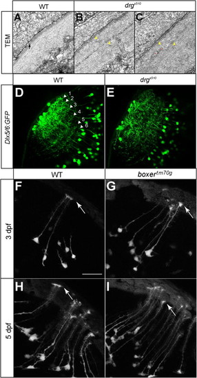
Lamination of Tectal Dendrites Is Disrupted in drgs510 Mutants and Radial Glia Appear Morphologically normal in box Mutants, Related to Figure 5(A–C) TEM photographs of the basement membrane of WT (A) and drgs510 (B, C) tectum at 5 dpf. The arrow in A points at the lamina densa in WT. Yellow arrowheads in B and C point at the putative lamina densa in drgs510 tectum(B, C). Instead of a smooth lamina densa, which contains type IV Collagen, two thin electron-dense layers are present in drgs510. Red stars indicate neurites, probably axonal profiles, aberrantly projecting into the ECM near the surface. Scale bar is 0.5 µm.(D and E) Single optical sections of Dlx5/6:GFP transgenic fish at 6 dpf. The WT pattern (D) shows six laminae. In drgs510 (F), these laminae are present but more diffuse, and the superficial-most terminations are disrupted.(F–I) Projections of radial glia in Gal4s1082t crossed to UAS:Kaede in WT (F, H) and boxtm70 g (G, I) at 3 dpf (F, G) and 5 dpf (H, I). Arrows point to glial endfeet. Radial glia and their endfeet appear normal in the mutant. Scale bar (in F, for panels F-I) is 10μm. |
Reprinted from Cell, 146(1), Xiao, T., Staub, W., Robles, E., Gosse, N.J., Cole, G.J., and Baier, H., Assembly of Lamina-Specific Neuronal Connections by Slit Bound to Type IV Collagen, 164-176, Copyright (2011) with permission from Elsevier. Full text @ Cell