
Fig. 2
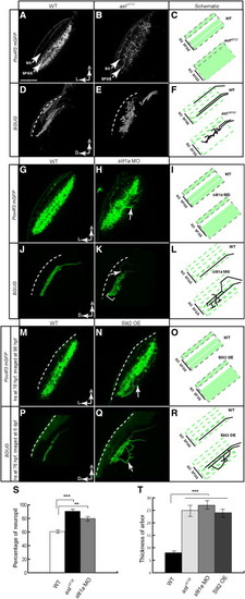
Robo/Slit Signaling Is Required for RGC Axon Laminar Targeting(A–C) Single optical sections of the tectum in Pou4f3:mGFP transgenic larvae at 6 dpf. RGC axons (labeled with membrane-targeted GFP) remain in their target lamina in the WT (A) and cross the laminar boundaries in the astti272z mutant (B). (C) is a schematic summary.(D–F) Projections of confocal images of a WT (D) and astti272z (E) tectum in BGUG transgenic larva at 6 dpf. Image stacks were rotated to best show laminae. Arrows point at the axons leaving their target layer. Dashed lines indicate the skin overlaying the tectum. (F) is a schematic summary.(G–I) Single optical sections of the tectum in Pou4f3:mGFP transgenic larvae at 5 dpf. (G) is a control fish. (H) is a slit1a morphant. (I) is a schematic summary.(J–L) Projections of confocal images of the control (J) and slit1a morphant (K) tectum in BGUG transgenic larva at 5 dpf. (L) is a schematic summary.(M–O) Single optical sections of the tectum in Pou4f3:mGFP transgenic larvae. (M) is a control fish. (N) is a hsp70l:slit2-GFP transgenic carrier. Both were heat shocked at 76 hpf. (O) is a schematic summary.(P–R) Projections of confocal images of the control (P) and hsp70l:slit2-GFP (Q) tectum in BGUG transgenic larva at 6 dpf. Arrows point at axons projecting deeper than SFGS. (R) is a schematic summary.(S) Quantification of Pou4f3:mGFP RGC laminar position.(T) Quantification of RGC arbor thickness.***p < 0.001, **p < 0.05. The scale bar represents 20 μm. Error bars represent the standard error of the mean (SEM). See alsoFigure S1 and Table S1. |
Reprinted from Cell, 146(1), Xiao, T., Staub, W., Robles, E., Gosse, N.J., Cole, G.J., and Baier, H., Assembly of Lamina-Specific Neuronal Connections by Slit Bound to Type IV Collagen, 164-176, Copyright (2011) with permission from Elsevier. Full text @ Cell