
Fig. 1
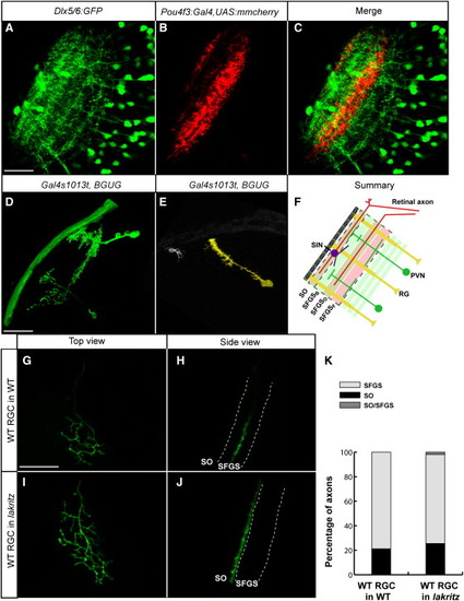
Axons, Dendrites, and Radial Glia Contribute to the Laminar Architecture of the Zebrafish TectumImages were taken at 6 dpf and are single optical sections (A–C) merged confocal Z stacks (projections) (D, E, and G–J).(A) Dlx5/6:GFP-labeled tectal neurons.(B) Pou4f3:Gal4 and UAS:mmCherry labeled RGC axons.(C) Merged image of (A) and (B).(D) Tectal neurons, sparsely labeled with Gal4s1013t and BGUG.(E) Radial glial cell and a superficial interneuron (SIN), labeled in the same tectum.(F) Schematic summary of the laminar composition of the larval tectum. Scale bars represent 20 μm.(G–J) Pou4f3:mGFP-positive, WT donor RGC transplanted into a nontransgenic host larva. Host was either the WT (G and H) or lakth241 (I and J). (G) and (I) are top views. (H) and (J) are rotated views of the confocal image stacks in (G) and (I) to best show axonal lamination.(K) Quantification of transplanted RGC laminar choices. Single axons behave very similarly in crowded (transplanted into the WT, n = 104) versus RGC-depleted (transplanted into lakth241, n = 52) conditions. |
Reprinted from Cell, 146(1), Xiao, T., Staub, W., Robles, E., Gosse, N.J., Cole, G.J., and Baier, H., Assembly of Lamina-Specific Neuronal Connections by Slit Bound to Type IV Collagen, 164-176, Copyright (2011) with permission from Elsevier. Full text @ Cell