
Fig. S6
- ID
- ZDB-FIG-110610-6
- Publication
- Wilkinson et al., 2009 - Hedgehog and Bmp polarize hematopoietic stem cell emergence in the zebrafish dorsal aorta
- Other Figures
- All Figure Page
- Back to All Figure Page
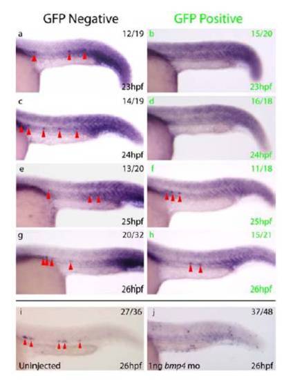
Expression of gfi1 Within the DA is Lost in a Similar Manner to runx1 Following Bmp Inhibition a-h) Expression of gfi1 (red arrowheads) in embryos heat shocked at 21hpf. Whereas gfi1 is expressed from 23-26hpf in two thirds of control GFP- (black letters) embryos, gfi1 is lost from the DA in 75% of GFP+ (green letters) embryos at 23hpf and 89% of GFP+ embryos at 24hpf, in a similar pattern to that observed for runx1 following induction of tBR at the same stages. i, j) Whilst gfi1 is expressed within the DA at 26hpf (red arrowheads) in 75% of uninjected controls (i), expression of gfi1 is absent from the DA in 77% of embryos injected with bmp4 morpholino (j). Taken together, these results indicate that temporal inhibition of Bmp signalling (a-h) results in temporal inhibition of gfi1 followed by recovery, but that permanent inhibition of Bmp via bmp4 morpholino injection results in a more permanent inhibition of gfi1 expression within the DA in a similar manner to that observed for runx1. Embryos were over stained to ensure that lack of gfi1 expression in the DA was not an artefact of reduced staining. |
Reprinted from Developmental Cell, 16(6), Wilkinson, R.N., Pouget, C., Gering, M., Russell, A.J., Davies, S.G., Kimelman, D., and Patient, R., Hedgehog and Bmp polarize hematopoietic stem cell emergence in the zebrafish dorsal aorta, 909-916, Copyright (2009) with permission from Elsevier. Full text @ Dev. Cell