
Fig. 2
- ID
- ZDB-FIG-090714-25
- Publication
- Wilkinson et al., 2009 - Hedgehog and Bmp polarize hematopoietic stem cell emergence in the zebrafish dorsal aorta
- Other Figures
- All Figure Page
- Back to All Figure Page
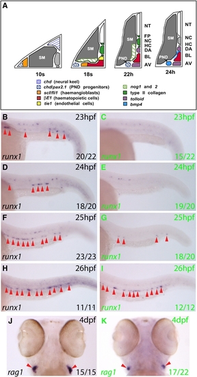
Bmp Signaling Is Required for Initiation of the HSC Program in the Ventral DA (A) Gene expression summary as progenitors migrate to the midline and form blood and DA/AV endothelium. The DA region becomes conducive to Bmp signaling by 24 hpf. (B–I) Expression of runx1 (arrows) in embryos heat shocked at 21 hpf. runx1 expression in the DA at 23–26 hpf (GFP-, black letters) was substantially reduced in GFP+ embryos (green letters) at 23–25 hpf. Representative embryos are depicted, and numbers with or without runx1 staining were scored. (J and K) rag1 expression in thymi at 4 dpf heat shocked at 21 hpf. 17/22 GFP+ embryos had reduced rag1 expression compared to GFP- embryos, in which rag1 staining showed little variation. The remaining 5/22 GFP+ embryos exhibited wild-type rag1 expression. AV, axial vein; BL, primitive blood; DA, dorsal aorta; HC, hypochord; NC, notochord; NT, neural tube; PND, pronephric duct; SM, somitic mesoderm. |
Reprinted from Developmental Cell, 16(6), Wilkinson, R.N., Pouget, C., Gering, M., Russell, A.J., Davies, S.G., Kimelman, D., and Patient, R., Hedgehog and Bmp polarize hematopoietic stem cell emergence in the zebrafish dorsal aorta, 909-916, Copyright (2009) with permission from Elsevier. Full text @ Dev. Cell