
Fig. S4
- ID
- ZDB-FIG-110610-4
- Publication
- Wilkinson et al., 2009 - Hedgehog and Bmp polarize hematopoietic stem cell emergence in the zebrafish dorsal aorta
- Other Figures
- All Figure Page
- Back to All Figure Page
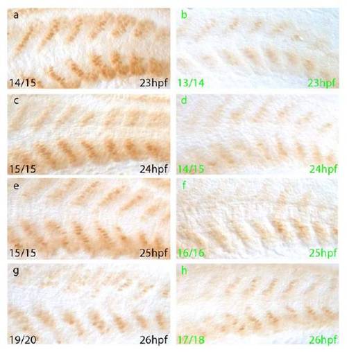
Heat Shock Induced Expression of tBR Leads to Reduced Levels of Phosphorylated Bmp Receptor Smads Whole-mount immunohistochemistry for phosphorylated Smad1, 5, 8 in 23-26hpf embryos from a tBR heterozygous outcross subjected to a 21hpf heat shock. a, c, e, g) Nuclear staining is clearly visible within the developing myotome of GFP negative embryos (black letters). b, d, f, h) Nuclear staining is substantially reduced in GFP positive embryos (green letters) at 23hpf, 24hpf and 25hpf in comparison to GFP negative embryos at the same stage. By 26hpf, differences in nuclear staining between GFP positive and GFP negative embryos are much less, indicating turnover of dominant negative receptor and recovery of Bmp signalling within the trunk to wild-type levels at this time. |
Reprinted from Developmental Cell, 16(6), Wilkinson, R.N., Pouget, C., Gering, M., Russell, A.J., Davies, S.G., Kimelman, D., and Patient, R., Hedgehog and Bmp polarize hematopoietic stem cell emergence in the zebrafish dorsal aorta, 909-916, Copyright (2009) with permission from Elsevier. Full text @ Dev. Cell