
Fig. 7
- ID
- ZDB-FIG-110516-62
- Publication
- Lauter et al., 2011 - Multicolor fluorescent in situ hybridization to define abutting and overlapping gene expression in the embryonic zebrafish brain
- Other Figures
- All Figure Page
- Back to All Figure Page
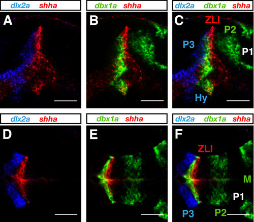
Three-color FISH. Embryos at 1 dpf were hybridized simultaneously with fluorescein-labeled dbx1a, dinitrophenyl-labeled shha and digoxigenin-labeled dlx2a RNA probes, sequentially detected and visualized using DyLight633-, TAMRA- and FAM-tyramide, respectively. (A-F) In lateral (A-C) and dorsal (D-F) views with anterior to the left, dbx1a, shha and dlx2a expression is shown in green, red and blue, respectively. Overlays of two (A, B, D, E) or all three (C, F) different channels of the same confocal plane are shown. Photographs were taken on a LSM510 confocal microscope (Carl Zeiss). Images were false-colored with RGB look-up tables and processed using ImageJ software. Scale bar = 50 μm. Hy, hypothalamus; M, midbrain; P1, P2, and P3, prosomeres 1, 2, and 3; ZLI, zona limitans intrathalamica. |