
Fig. 3
- ID
- ZDB-FIG-110516-58
- Publication
- Lauter et al., 2011 - Multicolor fluorescent in situ hybridization to define abutting and overlapping gene expression in the embryonic zebrafish brain
- Other Figures
- All Figure Page
- Back to All Figure Page
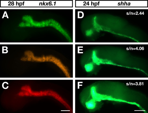
Bench-made fluorescent tyramides applied in zebrafish whole-mount FISH. Embryos were viewed on an Axioplan II microscope (Carl Zeiss). For excitation, a mercury burner (HBO 103 OSRAM) was used. Lateral views of zebrafish brains are shown with anterior to the left. Scale bar = 100 μm. (A-C) Zebrafish embryos at 28 hpf hybridized to a digoxigenin-labeled nkx6.1 antisense RNA probe. Images were captured with an Orca digital camera (Hamamatsu). As POD substrates, three different bench-made fluorogenic tyramides at a 1:250 dilution were used: (A) FAM-tyramide (exposure time, 100 ms; Chroma-filter 41001); (B) TAMRA-tyramide (exposure time, 4 ms; Chroma-filter 41002b); and (C) DyLight633-tyramide (exposure time, 120 ms; Chroma-filter 41008). (D-F) Zebrafish embryos at 24 hpf hybridized to a digoxigenin-labeled shha antisense RNA probe. Images were recorded with an Axiocam digital color camera (Carl Zeiss) using identical exposure times. Transcript distribution was visualized with (D) fluorescein-tyramide from Perkin Elmer (SAT701B001EA) at a 1:100 dilution, (E) bench-made FAM-tyramide at a 1:250 dilution, or (F) a 1:100 dilution. |