
Fig. 5
- ID
- ZDB-FIG-110516-60
- Publication
- Lauter et al., 2011 - Multicolor fluorescent in situ hybridization to define abutting and overlapping gene expression in the embryonic zebrafish brain
- Other Figures
- All Figure Page
- Back to All Figure Page
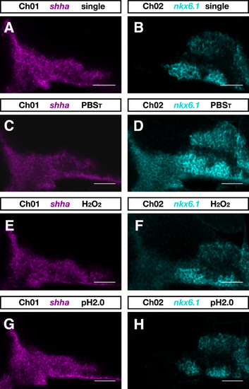
Inactivation of antibody-POD conjugate. Zebrafish embryos at 28 hpf were hybridized with dinitrophenyl-labeled shha and digoxigenin-labeled nkx6.1 RNA probes. (A, B) The expression patterns of shha (A) and nkx6.1 (B) as seen in single-color FISH experiments. In two-color experiments shha transcript was detected first using DyLight633-tyramide and nkx6.1 transcript was detected subsequently by FAM-tyramide. (C-H) Prior to the second round of detection, embryos were incubated for 10 minutes in PBST (PBS plus 0.1% Tween-20) (C, D), PBST containing 6% H2O2 (E, F), or 100 mM glycine-HCl pH 2.0 (G, H). Single confocal sections of zebrafish brains are shown in the DyLight633-detection channel (Ch01) and in the FAM-detection channel (Ch02) from a lateral view and with anterior to the left. Images were recorded on a LSM510 microscope (Carl Zeiss) and false colored in ImageJ. Scale bar = 50 μm. |