|
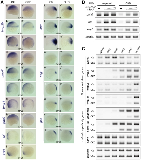
Defects in DV patterning and gastrulation movements in the QKD embryos. (A) DV patterning defects involving the reduced expression of bmp genes. Expression of bmp2b/7/4 (a–g) and the Bmp downstream genes gata2, szl and eve1 (h–j) is reduced in the QKD embryos. (k–s) Expression of the Bmp antagonist genes, chd and nog1, and the organizer gene gsc. All are lateral views with dorsal to the right. (B) The BMP downstream genes are restored by an exogenous supply of bmp2b/7. Embryos were injected together with the MOs for QKD and a mixture of bmp2b/7 mRNAs (20 or 40 pg each) and subjected to RT-PCR analysis at the shield stage. bactin1 was used as an RT-PCR control. (C) Decreased expression of genes regulating C&E movements in the QKD embryos. Temporal expression profiles of the indicated genes in the uninjected control and QKD embryos from the sphere to 3-somite stages were determined by RT-PCR. Expression of non-canonical wnt genes is reduced in the QKD embryos. Expression of pcdh18a/18b is also reduced in the QKD embryos, whereas cdh1, which is known to be involved in epiboly, is expressed at normal levels. bactin1 was used as an RT-PCR control.
|